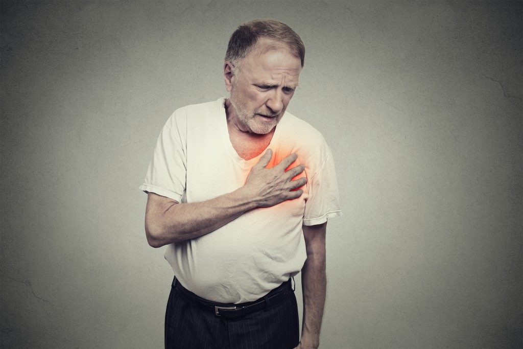
Cardiac conditions are, unfortunately, now among the leading causes of morbidity and mortality. Fortunately, the medical sector has come up with several ways of ensuring patients access the best healthcare when suffering from the same. The primary elements of optimal care for cardiac patients are regular monitoring of the heart and making an accurate diagnosis. While these were challenging in the past, they have been made possible and easy with imaging tests.
An ultrasound scan done at a London-based imaging center is among the baseline imaging studies for cardiac patients. Echocardiography nonetheless marks the most specific imaging test for the heart and uses sound waves for the production of real-time images of the heart. The images generated are known as echocardiograms. They help doctors spot blood clots in your cardiac vessels, fluid around the heart’s sac, and issues with the aorta. Echo scans can be ordered if you have signs of cardiac conditions like unexplained chest pain or shortness of breath and irregular heartbeat. Here are the types of echo scans that might be used.
Transthoracic Echocardiography
This common echo scan type is noninvasive and painless. In it, a transducer is placed on your ribcage over the heart. The device conveys ultrasound waves towards the heart through your chest. A computer linked to the device then displays the waves when they bounce to the transducer. The process generates a live image that can be interpreted to determine your cardiac functioning.
Transesophageal Echocardiography
This type is often ordered when the transthoracic echo scan does not generate definitive images. In this echocardiography, a small transducer is passed down your throat using a small flexible tube placed in your mouth. Your throat will be numbed before the tube’s insertion to minimize your discomfort. The tube travels down to your stomach while the transducer falls behind the heart and generates live images of its condition.
Three-Dimensional Echocardiography
This type creates 3D images of your cardiac vessels. It uses either of the previous ones to do it. The images used in this instance are multiple ones taken from different angles. 3D echo scans are often used before undergoing heart valve surgery. They nonetheless can also be used for the diagnosis of pediatric cardiac conditions.
Doppler Echocardiography
This test measures and assesses the blood flowing through your heart’s valves and chambers. The blood amount pumped with each heartbeat indicates your cardiac functioning. Doppler echocardiography can pick issues with your cardiac valves and walls or clots in the valves.
Fetal Echocardiography

This type is used in pregnancy at 18-22 weeks gestation. In the test, a transducer is positioned over the belly to detect cardiac issues in a fetus. Fetal echocardiography, like all echo scan types, uses no radiation and is thus safe for your unborn child.
There is generally no preparation needed before these echo scans. If, however, you are undergoing a transesophageal scan, you might be instructed to fast a few hours before the procedure to prevent vomiting. Also, you should notify the doctor if you are taking any medications or herbal supplements or have a pacemaker or medical condition.